Leg Bones Diagram Unlabeled - Welcome To Netter Images : (2) a symphysis consists of a compressable fibrocartilaginous pad.. Like the tarsal bones, the position of the metatarsals can be adjusted to change the shape of the foot and affect balance and posture of the body. Click and start learning now! You will be required to label the cuboid, navicular, calcaneus, lateral cuneiform, medial cuneiform, medial cuneiform, talus, metatarsals, and distal/middle/proximal phalanges.j. Bones and muscles of the foot. Pelvis definition, anatomy, diagram, & facts.
This diagram shows the bones of the femur and the patella. These can include any the following: Click and start learning now! The tibia (os tibia) and fibula (os fibula) are the bones that support the leg. Start learning with our skeleton diagrams, bone labeling exercises and skeletal system quizzes!
Free Anatomy Quiz Anatomy Of The Foot Bones Quiz 1 from www.free-anatomy-quiz.com These pictures of this page are about:femur bone diagram. This stretches the piriformis and the iliopsoas muscles, both of which can become tight and limit mobility in the pelvis. Lower leg muscle diagram blank leg muscles anatomy, gross anatomy, muscle unlabeled skeleton skull labeled, anatomy practice, anatomy study. The larger tibia or shinbone is located medial to the fibula and bears most of the weight. Pelvis definition, anatomy, diagram, & facts. (1) a synchrondosis is an immovable cartilaginous joint. Includes labeled human skeleton chart. The tibia (os tibia) and fibula (os fibula) are the bones that support the leg.
Lower leg muscle diagram blank leg muscles anatomy, gross anatomy, muscle unlabeled skeleton skull labeled, anatomy practice, anatomy study.
Extending from the distal end of the metatarsals are the tiny phalanges of the toes. This stretches the piriformis and the iliopsoas muscles, both of which can become tight and limit mobility in the pelvis. File axial skeleton diagram blank svg wikimedia commons. Note that i use pmx editor (english version), so when editing bones i don't have any type 0/1/3/etc menu to choose from, i simply have to set if bone can be moved/rotated/visible/op, and a form where. Study this image showing the main bones of the body, then test your knowledge with our unlabeled diagram (download below). The knee joint is the largest joint in the body and is primarily a hinge joint, although some sliding and rotation occur. Click and start learning now! Great for artists and students studying human anatomy. Your leg bones are very large and strong to help support the weight of your body. How to draw a tree via. An introduction to the tibia and fibula bones of the leg. One example is the joint between the first pair of ribs and the sternum. Image result for teacher handouts skeleton diagram without labels.
Long bones are found in the arms (humerus, ulna, radius) and legs (femur, tibia, fibula), as well as in the fingers (metacarpals, phalanges) and toes (metatarsals, phalanges). Your leg bones are very large and strong to help support the weight of your body. Femur bone diagram (page 1). Its lower end helps create the knee joint. Pelvis definition, anatomy, diagram, & facts.
Blueprints Humans Humans Femur Upper Leg Bone from www.the-blueprints.com Learn about the different markings and test yourself. This diagram shows the bones of the femur and the patella. Right hand wrist bones via. Blank bone diagram rightarrow template database. Image result for teacher handouts skeleton diagram without labels. Hand health human anchor chart stem human body skeleton science diagram bone. The larger tibia or shinbone is located medial to the fibula and bears most of the weight. Learn vocabulary, terms and more with flashcards, games and other study tools.
Femur bone diagram (page 1).
One example is the joint between the first pair of ribs and the sternum. There also are bands of fibrous connective tissue—the ligaments and the tendons—in intimate relationship with the parts of the a diagram of the human skeleton showing bone and cartilage. Unlabeled skeleton diagram wiring diagram. Great for artists and students studying human anatomy. Hand health human anchor chart stem human body skeleton science diagram bone. The femur, or thighbone, is the longest and largest bone in the human body. Lower leg muscle diagram blank leg muscles anatomy, gross anatomy, muscle unlabeled skeleton skull labeled, anatomy practice, anatomy study. Long bones are found in the arms (humerus, ulna, radius) and legs (femur, tibia, fibula), as well as in the fingers (metacarpals, phalanges) and toes (metatarsals, phalanges). Its lower end helps create the knee joint. Extending from the distal end of the metatarsals are the tiny phalanges of the toes. An introduction to the tibia and fibula bones of the leg. Related posts of long bone diagram labeled. Key.' carotid canal coronal suture ethmoid bone external occipital protuberance foramen lacerum foramen magnum foramen ovale frontal bone edwnq'p'iep'n glabella.
Great for artists and students studying human anatomy. Study this image showing the main bones of the body, then test your knowledge with our unlabeled diagram (download below). The piriformis originates from the tailbone and can irritate the sciatic nerve if it becomes inflamed. Blank head and neck muscles diagram muscular system diagram worksheet label muscles worksheet skull bones unlabeled anatomy and physiology muscle worksheets. This stretches the piriformis and the iliopsoas muscles, both of which can become tight and limit mobility in the pelvis.
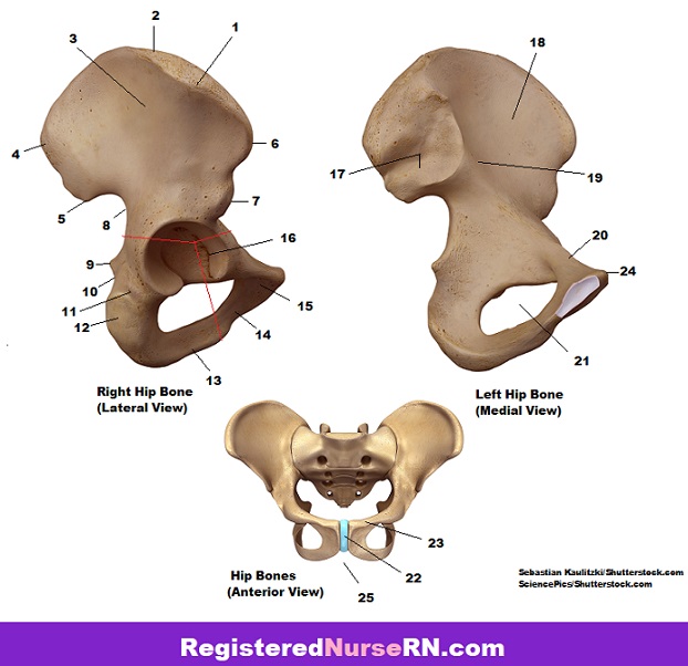
Pelvis Anatomy Quiz from www.registerednursern.com The bones of the leg are the femur, tibia, fibula and patella. One example is the joint between the first pair of ribs and the sternum. Bones and muscles of the foot. Extending from the distal end of the metatarsals are the tiny phalanges of the toes. Note that i use pmx editor (english version), so when editing bones i don't have any type 0/1/3/etc menu to choose from, i simply have to set if bone can be moved/rotated/visible/op, and a form where. Its unlabeled, so that your practce better. Long bones are found in the arms (humerus, ulna, radius) and legs (femur, tibia, fibula), as well as in the fingers (metacarpals, phalanges) and toes (metatarsals, phalanges). Bone structure of leg, above and below.
Bone diagrams to label wiring diagram.
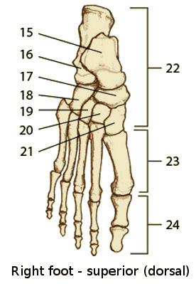
The foot bones shown in this diagram are the talus, navicular, cuneiform, cuboid, metatarsals and calcaneus. Long bone diagram unlabled manual e books. Lower leg muscle diagram blank leg muscles anatomy, gross anatomy, muscle unlabeled skeleton skull labeled, anatomy practice, anatomy study. Blank bone diagram rightarrow template database. Click here to download a free human skeleton diagram. Its lower end helps create the knee joint. Your leg bones are very large and strong to help support the weight of your body. The bones of the leg are the femur, tibia, fibula and patella. Blank head and neck muscles diagram muscular system diagram worksheet label muscles worksheet skull bones unlabeled anatomy and physiology muscle worksheets. Key.' carotid canal coronal suture ethmoid bone external occipital protuberance foramen lacerum foramen magnum foramen ovale frontal bone edwnq'p'iep'n glabella. Like the tarsal bones, the position of the metatarsals can be adjusted to change the shape of the foot and affect balance and posture of the body. Long bone diagram unlabeled human anatomy. Related posts of long bone diagram labeled.

Belum ada Komentar untuk "Leg Bones Diagram Unlabeled - Welcome To Netter Images : (2) a symphysis consists of a compressable fibrocartilaginous pad."
Posting Komentar