Muscles Of The Chest And Abdomen Labeled : Ventral view of the thorax skeleton. — anatomy references ... : Ventral neck, chest and abdomen:. The skeletal muscles of the abdomen form part of the abdominal wall, which holds and protects the gastrointestinal system. The muscle striations, are they easily visible on the cat as they are in the dissection book or are they procedure: The serratus anterior is located more laterally in the chest wall and forms the medial border of the axilla region. The muscles of the anterior abdominal wall are located near the midline between the costal margin superiorly and the pubis inferiorly. They are the pectoralis major, pectoralis minor, and the serratus anterior.
Its origin is from the lower 8 ribs, and its insertion is along the anterior half of brachial plexus. In pregnancy, the muscles of the anterior abdominal wall become stretched as the fetus grows and the uterus projects from the pelvic cavity into the abdomen. Cardiac muscle forms the heart and is not part of the musculoskeletal system. For some smaller muscle observations, larger. The abdomen (colloquially called the belly, tummy, midriff or stomach) is the part of the body between the thorax (chest) and pelvis, in humans and in other vertebrates.
Anatomy CT Axial Abdomen and Pelvis Male from 1.bp.blogspot.com Linea alba (white line of connective tissue at midline). What is this muscle?what is its origin and insertion?function? Muscle performance in neck pain assessment and rehab of the deep. Abdominal muscles help you breathe out when you are breathing fast, such as during physical activity. The serratus anterior is located more laterally in the chest wall and forms the medial border of the axilla region. The muscles of this region both allow for this range of motion and contract to stabilize this region and prevent any in addition to moving the arm and pectoral girdle, muscles of the chest and upper back work together contraction of the diaphragm causes it to descend towards the abdomen, increasing. The abdominal muscles stretch over the abdomen from the chest to the hips, covering the center and sides also. The pectoralis mayor works the most if you do push up or bench press like moves, but since you push, your deltoids and triceps work as well.
Related online courses on physioplus.
The abdominal muscles stretch over the abdomen from the chest to the hips, covering the center and sides also. Abdominal muscle, any of the muscles of the anterolateral walls of the abdominal cavity, composed of three flat muscular sheets, from without inward: The pectoralis major is located on the upper portion of the sternum and lies along most of the entire length of the humerus. The external oblique muscle is a broad muscle that runs along the anterolateral abdomen and chest wall. Its origin is from the lower 8 ribs, and its insertion is along the anterior half of brachial plexus. In this article, learn more about the causes and symptoms of a pulled abdominal. Muscle performance in neck pain online course: It works to move forelimb towards the chest. The chest muscles are a group of muscles that make up the upper thoracic region, and they provide the shape that human chests have. Muscular wall separating the chest and abdomen. For some smaller muscle observations, larger. The skeletal muscles of the abdomen form part of the abdominal wall, which holds and protects the gastrointestinal system. They are the pectoralis major, pectoralis minor, and the serratus anterior.
Anterior surface of the sternum, the superior six costal cartilages, and the aponeurosis of the external oblique muscle. The external oblique muscle is a broad muscle that runs along the anterolateral abdomen and chest wall. The primary function is certainly to provide support to the skeletal system and to facilitate its movements. The muscles of the chest are the following ones. Common chest and abdominal injuries.
Anterior View of the Muscles of the Trunk | ClipArt ETC from etc.usf.edu In pregnancy, the muscles of the anterior abdominal wall become stretched as the fetus grows and the uterus projects from the pelvic cavity into the abdomen. The muscles of the anterior abdominal wall are located near the midline between the costal margin superiorly and the pubis inferiorly. Human anatomy hd pic abdominal 12 photos of the human anatomy hd pic abdominal , human muscles. Abdominal muscle, any of the muscles of the anterolateral walls of the abdominal cavity, composed of three flat muscular sheets, from without inward: As the abdominal muscles are hard to support externally, treatment involves rest and pain medication. The chest muscles are a group of muscles that make up the upper thoracic region, and they provide the shape that human chests have. Labeling muscles (chest and abdomen). Muscle performance in neck pain online course:
For some smaller muscle observations, larger.
As the abdominal muscles are hard to support externally, treatment involves rest and pain medication. Related online courses on physioplus. Like skeletal muscle, cardiac muscle has a regular pattern of fibers that also appear as stripes under a microscope. How to build ab and chest. The muscles of the chest are the following ones. The serratus anterior is located more laterally in the chest wall and forms the medial border of the axilla region. This causes air to flow in. Abdominal muscle, any of the muscles of the anterolateral walls of the abdominal cavity, composed of three flat muscular sheets, from without inward: They are the pectoralis major, pectoralis minor, and the serratus anterior. However, cardiac muscle contracts and relaxes rhythmically without a person's awareness. The pectoralis mayor works the most if you do push up or bench press like moves, but since you push, your deltoids and triceps work as well. The pectoantebrachialis has been separated from the underlying pectoralis major, and is being lifted in the image. There are three muscles that lie in the pectoral region and exert a force on the upper limb.
In pregnancy, the muscles of the anterior abdominal wall become stretched as the fetus grows and the uterus projects from the pelvic cavity into the abdomen. Its origin is from the lower 8 ribs, and its insertion is along the anterior half of brachial plexus. The pectoantebrachialis has been separated from the underlying pectoralis major, and is being lifted in the image. The external oblique muscle is a broad muscle that runs along the anterolateral abdomen and chest wall. Ventral neck, chest and abdomen:
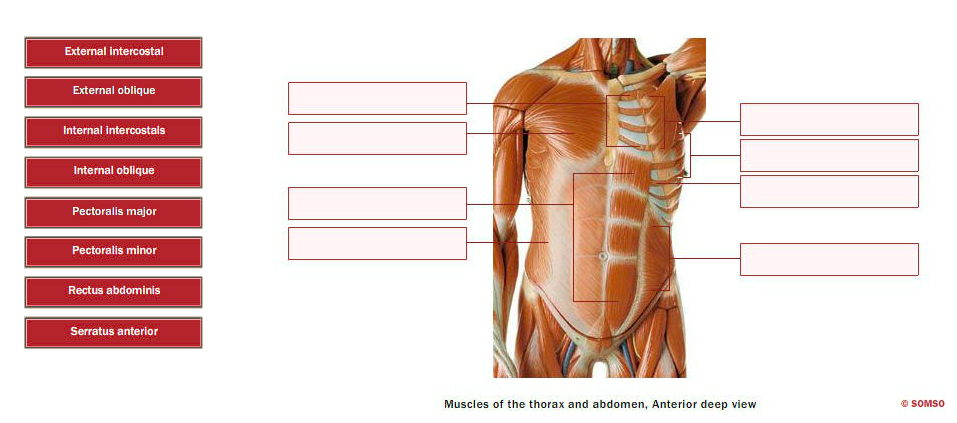
Solved: External Intercostal External Oblique Internal Int ... from media.cheggcdn.com We began our journey towards the muscles today. Fabian identifying the muscles and landmarks of the abdomen and chest. Common chest and abdominal injuries. The pectoantebrachialis has been separated from the underlying pectoralis major, and is being lifted in the image. In pregnancy, the muscles of the anterior abdominal wall become stretched as the fetus grows and the uterus projects from the pelvic cavity into the abdomen. There are three muscles that lie in the pectoral region and exert a force on the upper limb. Linea alba (white line of connective tissue at midline). The muscles of the chest are the following ones.
The skeletal muscles of the abdomen form part of the abdominal wall, which holds and protects the gastrointestinal system.
How do the cat muscles look the goal or procedure for this part was to examine the chest and abdomen. Muscle of the frog by label. The muscle striations, are they easily visible on the cat as they are in the dissection book or are they procedure: Abdominal muscles help you breathe out when you are breathing fast, such as during physical activity. Home › create › flashcards › health › abdomen › abdomen and chest muscles. As the abdominal muscles are hard to support externally, treatment involves rest and pain medication. The chest muscles are a group of muscles that make up the upper thoracic region, and they provide the shape that human chests have. The muscles of the chest are the following ones. Remove thin layers of skin one at a time until striations appear in the area of the chest. Muscle performance in neck pain online course: This causes air to flow in. Muscular wall separating the chest and abdomen. They are the pectoralis major, pectoralis minor, and the serratus anterior.
Home › create › flashcards › health › abdomen › abdomen and chest muscles muscles of the chest abdomen. Cardiac muscle forms the heart and is not part of the musculoskeletal system.
Belum ada Komentar untuk "Muscles Of The Chest And Abdomen Labeled : Ventral view of the thorax skeleton. — anatomy references ... : Ventral neck, chest and abdomen:"


Belum ada Komentar untuk "Muscles Of The Chest And Abdomen Labeled : Ventral view of the thorax skeleton. — anatomy references ... : Ventral neck, chest and abdomen:"
Posting Komentar